Forget the basic eye chart. Modern eye care delves deeper, mapping the intricate landscape of your cornea with precision. This is about understanding the very foundation of your vision.
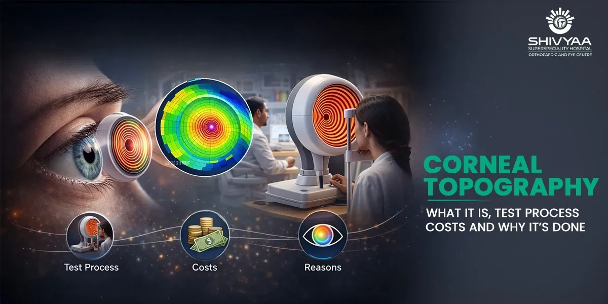
Corneal topography is a critical diagnostic tool. Think of it as creating a detailed 3D elevation map of your eye’s clear front surface. This map reveals subtle curvatures, irregularities, and power distributions that standard tests miss.
We use it for everything from fitting specialized contact lenses to planning life-changing laser vision correction. It’s the gold standard for detecting conditions like keratoconus long before they significantly impact sight.
Let’s explore how this technology works, why you might need it, and how it provides a roadmap for optimal eye health.
What is Corneal Topography?
Corneal topography is a painless and rapid scan that forms a detailed 3-D image of the cornea surface. The approximate time is five minutes, and reveals the little hills and valleys with which we see.
Imagine a map of a mountain range, but in your eye. The map has warm colors in steep areas and cool colours in flat ones. It is possible to describe an irregular pattern as what may have led to you seeing starbursts when nighttime comes, and your vision is 20/20.
Uses of Corneal Topography in Modern Eye Care
Corneal topography is your eye doctor’s high-precision GPS. It provides the navigational data required for complex procedures and accurate diagnoses. This single cornea test shifts treatment from estimation to exact planning.
We rely on corneal topography in three core clinical areas where standard measurements fail.
1. Planning and Screening for Vision Correction Surgery
Any laser treatment involves a close examination of the cornea by surgeons using a detailed map of the cornea. The map reveals the cornea’s shape and stability, and also examines the hidden irregularities like forme fruste keratoconus that may result in complications following LASIK. This data informs the surgery plan, particularly in topography-guided surgery, which corrects abnormal shapes of the cornea.
2. Fitting Specialty Contact Lenses
When standard soft lenses cause discomfort or poor vision, the problem is often corneal shape.
Corneal topography provides the blueprint for fitting:
- Rigid Gas Permeable (RGP) lenses.
- Scleral lenses that vault the entire cornea.
- Hybrid lenses
The accurate map makes sure that the lens fits perfectly on the surface of your eye. It removes guesswork, enhances the stability of the lens, enhances oxygen circulation, and maximizes comfort.
3. Diagnosing and Managing Corneal Diseases
This is where the technology proves indispensable. Corneal topography tracks minute changes over time, offering an objective baseline. We use it to:
- Diagnose and follow the course of Keratoconus.
- Evaluate the effects of dystrophies or corneal scars.
- Assess healing and astigmatism of transplant or injury.
One scan will show a pattern of a disease; however, a comparison of scans over the years reveals the real history of a disease that is either stable or progressive.
The Corneal Topography Experience: A Walkthrough From Prep to Results
Worried about the scan? Don’t be. The normal corneal topography test is one of the easiest and fastest diagnostics in the clinic. You’ll spend more time in the waiting room than in the chair. Here’s exactly what happens, from preparation to reading your map.
Preparing for Your Corneal Topography Test
Proper preparation ensures the most accurate mapping of your cornea’s natural shape. Follow these steps to avoid skewed results.
- Discontinue Contact Lenses: This is essential. Soft lenses should be removed at least 48-72 hours prior. Hard (RGP or scleral) lenses need a longer hiatus (usually two weeks or more) due to their ability to temporarily remodel the cornea.
- Arrive Makeup-Free: Do not use eye creams, lotion, or mascara on the day you are taking your test. The imaging may be disturbed by residue.
- Bring Your Prescriptions: Have your current eyeglasses and contact lens information available. This helps your doctor correlate your vision correction with the corneal map.
Clinical Insight: The most common reason for an unreliable scan is insufficient time out of contact lenses, especially rigid ones. We need to see your cornea’s true shape, not the temporary mold created by the lens.
During the Corneal Topography Procedure
The scan itself is quick and comfortable. In front of the topographer – a machine resembling an oversized digital camera, you will sit. Each eye takes only a few seconds to do the process.
- Positioning: You will have your chin and forehead on supports so that your head is still.
- Imaging: The technician will request that you concentrate on a central light. This machine displays a pattern of lit rings in your eye and captures a few digital images within a few seconds.
- Sensation: You will enjoy a light that has no flash. Nothing touches your eye. The entire process is not active on your part and requires an average duration of less than ten minutes to complete on both eyes.
Interpreting Your Corneal Topography Results
Your doctor will analyze a series of color-coded maps generated by the scan. These are not photographs but detailed topographic representations.
- Understanding the Map Colors: The system uses a consistent scale. Reds and oranges indicate steeper areas of the cornea with greater refractive power. Blues and greens represent flatter areas. A balanced, symmetrical pattern is typical of a normal, healthy corneal shape.
- Key Patterns We Assess: We evaluate the map for symmetry, smoothness, and the pattern of curvature. For example, a localized steep area in the inferior cornea may suggest early keratoconus. Irregular, asymmetric patterns can explain symptoms like glare or ghosting that aren’t correctable with standard glasses.
Corneal Topography Cost in India
Knowing the price of a corneal topography test will enable you to make a budget and not get caught on a bad foot. Prices are quite different depending on the place as well as the technology in India. This diagnostic scan will cost you an average of 1,500 to 7,000.
What Influences the Final Price?
The final invoice depends on more than just the machine. Consider these variables when requesting a quote.
- Technology Tier: Intrinsic Placido-disk topographers are affordable. Higher systems, including the pentacam which maps the front and back side are more expensive; the scan may cost 2000-5000 rupees.
- Clinical Setting & Location: A renowned specialty eye hospital in Mumbai or Ahmedabad will have different pricing than a standalone optometry practice in a tier-2 city. Reputation and overhead costs factor in.
- Purpose of the Test: Is it a standalone diagnostic? Or is it bundled into a pre-surgical package for LASIK or ICL? Bundled pricing often offers better value but may not itemize the topography cost separately.
Who Really Needs a Corneal Topography Test?
Consider corneal topography a diagnostic measure, not a screening. We would recommend using it in special and complicated questions concerning corneal health and vision. The scan is necessary in case of falling into one of the categories below.
1. Refractive Surgery Candidates (LASIK, PRK, Contoura Vision)
Corneal topography is a must if you intend to undergo laser vision correction. Your pre-operative workup relies on it. The scan eliminates any undetectable contraindications, such as early keratoconus, and gives a more exact corneal reading to be used in setting up the laser to treat in a customized manner. Failure to scan aggravates the risk of surgery.
2. Patients with Known or Suspected Corneal Disease
This is a diagnostic that is critical in the treatment of corneal conditions. We use it to:
- Diagnosis and stage of keratoconus; the typical steep and inferior cone pattern is observed in the map.
- Track disease progression, e.g., pellucid marginal degeneration, corneal ectasia.
- Determine the effect of scars, dystrophies, or postinjury distortions on visual potential.
3. Challenging Contact Lens Fits
Corneal shape is frequently the culprit when regular soft lenses cannot perform their tasks; that is, they cause discomfort to the user, give them poor vision, or continuously pop out. Topography gives the plan on how to fit the advanced lenses:
- Rigid Gas Permeable (RGP) lenses
- Scleral lenses
- Custom hybrid designs
The lens will fit your individual corneal landmarks without guesswork as the map makes sure the lens fits.
Conclusion
The last, most important procedure before any vision correction procedure is learning your corneal topography. The blueprint is what provides safety, individualized treatment, and anticipates the most desirable outcome.
In Shivyaa Super Speciality Hospital, with the professional guidance of Dr. Yatri Bhavsar, our team employs accurate mapping to:
- Protect surgical safety, eliminate comorbidities.
- Develop completely tailored interventions of LASIK, Contoura Vision, et cetera.
- Corneal health and complex contact lens fits.
Don’t leave your vision to chance. Believe in the technology and skill that envisages each detail. Book your full corneal check-up with Shivyaa Hospital and be sure of the next step to clarity with all the confidence.